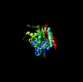
- The Protein Data Bank (PDB) is a repository for the 3-D structural data of large biological molecules, such as proteins and nucleic acids. (See also crystallographic database). The data, typically obtained by X-ray crystallography or NMR spectroscopy and submitted by biologistsbiochemists from around the world, are freely accessible on the Internet via the websites of its member organisations (PDBe, PDBj, and RCSB). The PDB is overseen by an organization called the Worldwide Protein Data Bank, wwPDB.
and The PDB is a key resource in areas of structural biology, such as structural genomics. Most major scientific journals, and some funding agencies, such as the NIH in the USA, now require scientists to submit their structure data to the PDB. If the contents of the PDB are thought of as primary data, then there are hundreds of derived (i.e., secondary) databases that categorize the data differently. For example, both SCOP and CATH categorize structures according to type of structure and assumed evolutionary relations; GO categorize structures based on genes.[1]
The PDB originated as a grassroots effort.[1] In 1971, Walter Hamilton of the Brookhaven National Laboratory agreed to set up the data bank at Brookhaven. Upon Hamilton's death in 1973, Tom Koeztle took over direction of the PDB. In January 1994, Joel Sussman was appointed head of the PDB. In October 1998,[2] the PDB was transferred to the Research Collaboratory for Structural Bioinformatics (RCSB); the transfer was completed in June 1999. The new director was Helen M. Berman of Rutgers University (one of the member institutions of the RCSB).[3] In 2003, with the formation of the wwPDB, the PDB became an international organization. The founding members are PDBe (Europe), RCSB(USA), and PDBj (Japan). The BMRB joined in 2006. Each of the four members of wwPDB can act as deposition, data processing and distribution centers for PDB data. The data processing refers to the fact that wwPDB staff review and annotates each submitted entry. The data are then automatically checked for plausibility. (The source code for this validation software has been made available to the public at no charge.
Gene Ontology
The Gene Ontology project provides an ontology of defined terms representing gene product properties. The ontology covers three domains:
The GO ontology is not static, and additions, corrections and alterations are suggested by, and solicited from, members of the research and annotation communities, as well as by those directly involved in the GO project. For example, an annotator may request a specific term to represent a metabolic pathway, or a section of the ontology may be revised with the help of community experts (e.g.[2]). Suggested edits are reviewed by the ontology editors, and implemented where appropriate.
The GO ontology file is freely available from the GO website in a number of formats, or can be accessed online using the GO browser AmiGO. The Gene Ontology project also provides downloadable mappings of its terms to other classification systems.
Enzyme Commission
The Enzyme Commission number (EC number) is a numerical classification scheme for enzymes, based on the chemical reactions they catalyze.[1] As a system of enzyme nomenclature, every EC number is associated with a recommended name for the respective enzyme.
Strictly speaking, EC numbers do not specify enzymes, but enzyme-catalyzed reactions. If different enzymes (for instance from different organisms) catalyze the same reaction, then they receive the same EC number.[2] By contrast, UniProt identifiers uniquely specify a protein by its amino acid sequence
KEGG Pathway
Type of protein
The Gene Ontology project provides an ontology of defined terms representing gene product properties. The ontology covers three domains:
- cellular component, the parts of a cell or its extracellular environment;
- molecular function, the elemental activities of a gene product at the molecular level, such as binding or catalysis;
- biological process, operations or sets of molecular events with a defined beginning and end, pertinent to the functioning of integrated living units: cells, tissues, organs, and organisms.
The GO ontology is not static, and additions, corrections and alterations are suggested by, and solicited from, members of the research and annotation communities, as well as by those directly involved in the GO project. For example, an annotator may request a specific term to represent a metabolic pathway, or a section of the ontology may be revised with the help of community experts (e.g.[2]). Suggested edits are reviewed by the ontology editors, and implemented where appropriate.
The GO ontology file is freely available from the GO website in a number of formats, or can be accessed online using the GO browser AmiGO. The Gene Ontology project also provides downloadable mappings of its terms to other classification systems.
Enzyme Commission
The Enzyme Commission number (EC number) is a numerical classification scheme for enzymes, based on the chemical reactions they catalyze.[1] As a system of enzyme nomenclature, every EC number is associated with a recommended name for the respective enzyme.
Strictly speaking, EC numbers do not specify enzymes, but enzyme-catalyzed reactions. If different enzymes (for instance from different organisms) catalyze the same reaction, then they receive the same EC number.[2] By contrast, UniProt identifiers uniquely specify a protein by its amino acid sequence
KEGG Pathway
KEGG PATHWAY is a collection of manually drawn pathway maps (see new maps, change history, and last updates) representing our knowledge on the molecular interaction and reaction networks for: 0. Global Map 1. Metabolism Carbohydrate Energy Lipid Nucleotide Amino acid Other amino acid Glycan Cofactor/vitamin Terpenoid/PK Other secondary metabolite Xenobiotics Overview 2. Genetic Information Processing 3. Environmental Information Processing 4. Cellular Processes 5. Organismal Systems 6. Human Diseases and also on the structure relationships (KEGG drug structure maps) in: 7. Drug Development | ||
- FtsH peptidase
Pseudomonas aeruginosa is an opportunistic Gram-negative pathogen that causes nosocomial infections for which there are limited treatment options. Penicillin-binding protein PBP3, a key therapeutic target, is an essential enzyme responsible for the final steps of peptidoglycan synthesis and is covalently inactivated by ?-lactam antibiotics. Here we disclose the first high resolution cocrystal structures of the P. aeruginosa PBP3 with both novel and marketed ?-lactams. These structures reveal a conformational rearrangement of Tyr532 and Phe533 and a ligand-induced conformational change of Tyr409 and Arg489. The well-known affinity of the monobactam aztreonam for P. aeruginosa PBP3 is due to a distinct hydrophobic aromatic wall composed of Tyr503, Tyr532, and Phe533 interacting with the gem-dimethyl group. The structure of MC-1, a new siderophore-conjugated monocarbam complexed with PBP3 provides molecular insights for lead optimization. Importantly, we have identified a novel conformation that is distinct to the high-molecular-weight class B PBP subfamily, which is identifiable by common features such as a hydrophobic aromatic wall formed by Tyr503, Tyr532, and Phe533 and the structural flexibility of Tyr409 flanked by two glycine residues. This is also the first example of a siderophore-conjugated triazolone-linked monocarbam complexed with any PBP. Energetic analysis of tightly and loosely held computed hydration sites indicates protein desolvation effects contribute significantly to PBP3 binding, and analysis of hydration site energies allows rank ordering of the second-order acylation rate constants. Taken together, these structural, biochemical, and computational studies provide a molecular basis for recognition of P. aeruginosa PBP3 and open avenues for future design of inhibitors of this class of PBPs.
| Authors: | |||
| Release Date: | 2010-12-22 | Classification: | Hydrolase/antibiotic |
| Experiment: | X-RAY DIFFRACTION with resolution of 2.00 Å | ||
| Compound: | 1 Polymer [ Display Full Polymer Details | Display for All Results ] | ||
| Citation: | Structural basis for effectiveness of siderophore-conjugated monocarbams against clinically relevant strains of Pseudomonas aeruginosa. (2010) Proc.Natl.Acad.Sci.USA [ Display Full Abstract | Display for All Results ] | ||
- Thermolysin
Thermolysin EC 3.4.24.27 is a thermostable neutral metalloproteinase enzyme produced by the gram-positive bacteria Bacillus thermoproteolyticus.[2] It requires one zinc ion for enzyme activity and four calcium ions for structural stability.[3] Thermolysin specifically catalyzes the hydrolysis of peptide bonds containing hydrophobic amino acids. However thermolysin is also widely used for peptide bond formation through the reverse reaction of hydrolysis.[4] Thermolysin is the most stable member of a family of metalloproteinases produced by various Bacillus species. These enzymes are also termed 'neutral' proteinases or thermolysin -like proteinases (TLPs).
Like all bacterial extracellular proteases thermolysin is first synthesised by the bacterium as a pre-proenzyme.[5] Thermolysin is synthesized as a pre-proenzyme consisting of a signal peptide 28 amino acids long, a pro-peptide 204 amino acids long and the mature enzyme itself 316 amino acids in length. The signal peptide acts as a signal for translocation of pre-prothermolysin to the bacterial cytoplasmic membrane. In the periplasm pre-prothermolysin is then processed into prothermolysin by a signal peptidase. The prosequence then acts as a molecular chaperone and leads to autocleavage of the peptide bond linking pro and mature sequences. The mature protein is then secreted into the extracellular medium.[6]
Thermolysin has a molecular weight of 34,600 Da. Its overall structure consists of two roughly spherical domains with a deep cleft running across the middle of the molecule separating the two domains. The secondary structure of each domain is quite different, the N-terminal domain consists of mostly beta pleated sheet, while the C-terminal domain is mostly alpha helical in structure. These two domains are connected by a central alpha helix, spanning amino acids 137-151.[7]
Thermolysin has a molecular weight of 34,600 Da. Its overall structure consists of two roughly spherical domains with a deep cleft running across the middle of the molecule separating the two domains. The secondary structure of each domain is quite different, the N-terminal domain consists of mostly beta pleated sheet, while the C-terminal domain is mostly alpha helical in structure. These two domains are connected by a central alpha helix, spanning amino acids 137-151.[7]
Structure
In contrast to many proteins that undergo conformational changes upon heating and denaturation, thermolysin does not undergo any major conformational changes until at least 70 °C.[8] The thermal stability of members of the TLP family is measured in terms of a T50 temperature. At this temperature incubation for 30 minutes reduces the enzymes activity by half. Thermolysin has a T50 value of 86.9 °C, making it the most thermo stable member of the TLP family.[9] Studies on the contribution of calcium to thermolysin stability have shown that upon thermal inactivation a single calcium ion is released from the molecule.[10] Preventing this calcium from originally binding to the molecule by mutation of its binding site, reduced thermolysin stability by 7 °C. However, while calcium binding makes a significant contribution to stabilising thermolysin, more crucial to stability is a small cluster of N-terminal domain amino acids located at the proteins surface.[9] In particular a phenylalanine (F) at amino acid position 63 and a proline (P) at amino acid position 69 contribute significantly to thermolysin stability. Changing these amino acids to threonine (T) and alanine (A) respectively in a less stable thermolysin-like proteinase produced by Bacillus stearothermophillus (TLP-ste), results in individual reductions in stability of 7 °C (F63→T) and 6.3 °C (P69→A) and when combined a reduction in stability of 12.3 °C.[9]Application
In the synthesis of aspartame, less bitter-tasting byproduct is produced when the reaction is catalyzed by thermolysin.[11]| Authors: | ||||||||||||||||||||||||
| Release Date: | 2010-12-08 | Classification: | Hydrolase | |||||||||||||||||||||
| Experiment: | X-RAY DIFFRACTION with resolution of 2.20 Å | |||||||||||||||||||||||
| Compound: | 1 Polymer [ Display Full Polymer Details | Display for All Results ] 2 Ligands [ Display Full Ligand Details | Display for All Results ] | |||||||||||||||||||||||
| Citation: | Not Available. | |||||||||||||||||||||||
- Leucyl Aminopeptidase
Leucyl aminopeptidases (or leucine aminopeptidases, LAPs) are enzymes that preferentially catalyze the hydrolysis of leucine residues at the N-terminus of peptides and proteins. Other N-terminal residues can also be cleaved, however. LAPs have been found across superkingdoms. Identified LAPs include bovine lens LAP, porcine LAP, Escherichia coli (E. coli) LAP (also known as PepA or XerB), and the solanaceous-specific acidic LAP (LAP-A) in tomato (Solanum lycopersicum).
Enzyme Description, Structure, and Active Site
The active sites in PepA and in bovine lens LAP have been found to be similar.[1] Shown in the picture below is the proposed model for the active site of LAP-A in tomato based on the work of Strater et al.[2][3]. It is also known that the
biochemistry of the LAPs from these three kingdoms is very similar. PepA, bovine lens LAP, and LAP-A preferentially cleave N-terminal leucine, arginine, and methionine residues. These enzymes are all metallopeptidases requiring divalent metal cations for their enzymatic activity [4] Enzymes are active in the presence of Mn+2, Mg+2 and Zn+2. These enzymes are also known to have high pH (pH 8) and temperature optima. At pH 8, the highest enzymatic activity is seen at 60oC. PepA, bovine lens LAP and LAP-A are also known to form hexamers in vivo. The Gu et al. from 1999 demonstrated that six 55kDA enzymatically inactive LAP-A protomers come together to form the 353kDa bioactive LAP-A hexamer. Structures of the bovine lens LAP protomer and the biologically active hexamer have been constructed [5] can be found through Protein Data Bank (2J9A).
| Authors: | |||
| Release Date: | 2010-09-08 | Classification: | Hydrolase |
| Experiment: | X-RAY DIFFRACTION with resolution of 2.60 Å | ||
| Compound: | 1 Polymer [ Display Full Polymer Details | Display for All Results ] 3 Ligands [ Display Full Ligand Details | Display for All Results ] | ||
| Citation: | Not Available. | ||


No comments:
Post a Comment